New Imaging Technology for Ultrahigh-Speed Brain Mapping
Brain structure and function have always been one of the important subjects in life science research. 3D microscopic imaging of brain structure is the basic method to understand how the brain works, yet the traditional 2D slice imaging technology makes brain structure reconstruction and analysis a tedious task.
Recently, a technique developed by professor BI Guoqiang and his team at the Center for Integrative Imaging (CII) of the school of life sciences at the University of Science and Technology of China (USTC) breaks that bottleneck.
Volumetric Imaging with Synchronized on-the-fly-scan and Readout (VISoR), the proposed method, has greatly improved the efficiency of brain imaging, with a series of new features such as large sample size, ultra-high speed, high resolution, avoiding motion blur and scalability.

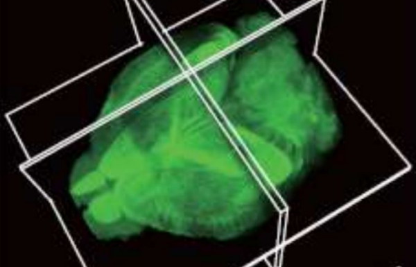
Whole brain VISoR imaging of a transgenic mouse. (Image by BI Guoqiang)
New technologies such as synchronized on-the-fly-scan and readout, combining synchronized scanning beam illumination and oblique imaging, etc. help VISoR obtain the imaging of large sample, such as mouse brain and monkey brain, with synaptic resolution efficiently and accurately.
This technique not only promotes the study of brain mapping, but also has a broad application prospects on other life science imaging applications, high-throughput pathological analysis and other medical fields.
The results were published in the National Science Review journal.
This research is one of the important research achievements of CII in recent years. CII is an innovative research and public technology platform established by the national research center for micro-scale material science of Hefei and the school of life sciences of USTC to promote the development of interdisciplinary research.
(USTC News Center, edited by HU Dongyin)
Back
